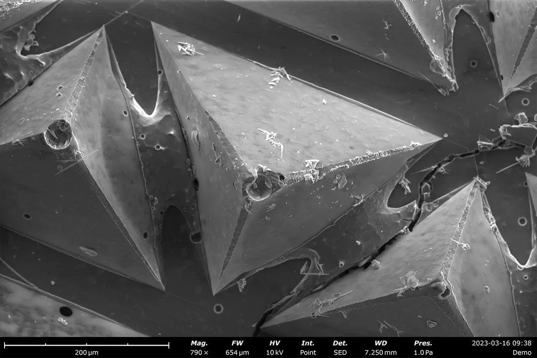
In a TEM image, what causes specific areas of the sample cross-section to appear darker?
Answer
Areas that are denser or absorb a greater number of electrons.
As the electron beam passes through the sample for TEM imaging, regions that are physically denser or absorb more electrons block more beam current, resulting in darker areas on the final image.
Related Questions
What fundamental physical limitation do electron microscopes bypass that constrains traditional light microscopes?What is the direct physical reason for the superior resolving power of an electron microscope compared to a light microscope?What is the typical maximum magnification limit for conventional light microscopes?What specific cellular components can electron microscopes visualize in extraordinary detail?What preparation step is typically necessary for biological samples in most electron microscopy, limiting observation of living processes?What is the primary function and resulting image characteristic of Scanning Electron Microscopy (SEM)?How is an image generated using Transmission Electron Microscopy (TEM)?In a TEM image, what causes specific areas of the sample cross-section to appear darker?Beyond biology, what structural element relevant to physical properties can electron microscopy reveal in materials science?What functional information about a material can be mapped simultaneously with structure using techniques like Cathodoluminescence Spectroscopy (CL)?