What do electron microscopes allow us to see?
The ability to peer into the minute architecture of the universe, long hidden from our naked eye, rests largely on the ingenious adaptation of electron microscopy. These instruments open up realms of observation fundamentally inaccessible to traditional light microscopes, revealing structures ranging from the fine details within a biological cell to the arrangement of atoms in a solid material. [6][7] Where conventional light microscopes, which use visible light, struggle to resolve objects smaller than about , [3] electron microscopes bypass this physical limitation entirely. [8]
# Resolution Limits
The core advantage of the electron microscope stems from the nature of the illumination source it employs. Standard optical microscopes are constrained by the wavelength of light itself; you cannot resolve objects smaller than roughly half the wavelength of the light used to view them. [3][8] Electrons, however, can be accelerated to extremely high speeds, resulting in a much, much shorter effective wavelength than that of visible light. [8] This dramatic reduction in wavelength translates directly into superior resolving power. [3] While light microscopes typically max out at magnifications around or , [9] electron microscopes routinely achieve magnifications far exceeding this, allowing researchers to see details that approach the atomic scale. [1][7] This shift in physical mechanism allows us to see the true complexity lurking beneath the cellular surface. [5]
# Cell Detail
In the realm of biology, electron microscopes permit us to see structures that are simply invisible using light-based methods. [6] This means moving past the general shape of a cell, which is visible with light microscopy, and into the precise organization of its internal machinery. [9] We can visualize the intricate network of organelles—the mitochondria, endoplasmic reticulum, and Golgi apparatus—in extraordinary detail. [2][6] Furthermore, these instruments are essential for studying entities much smaller than a full cell, such as individual viruses [2][6] and complex macromolecules. [2] The imaging reveals not just the presence of these components, but their morphology and spatial relationships within the tissue or cell matrix. [2] It is crucial to remember, though, that because most electron microscopy requires samples to be prepared in a vacuum to prevent the electron beam from scattering, the structures we observe are typically chemically fixed, dehydrated, and stained snapshots of life, rather than dynamic processes occurring in a living system. [1]
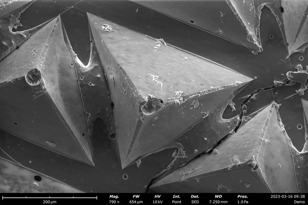
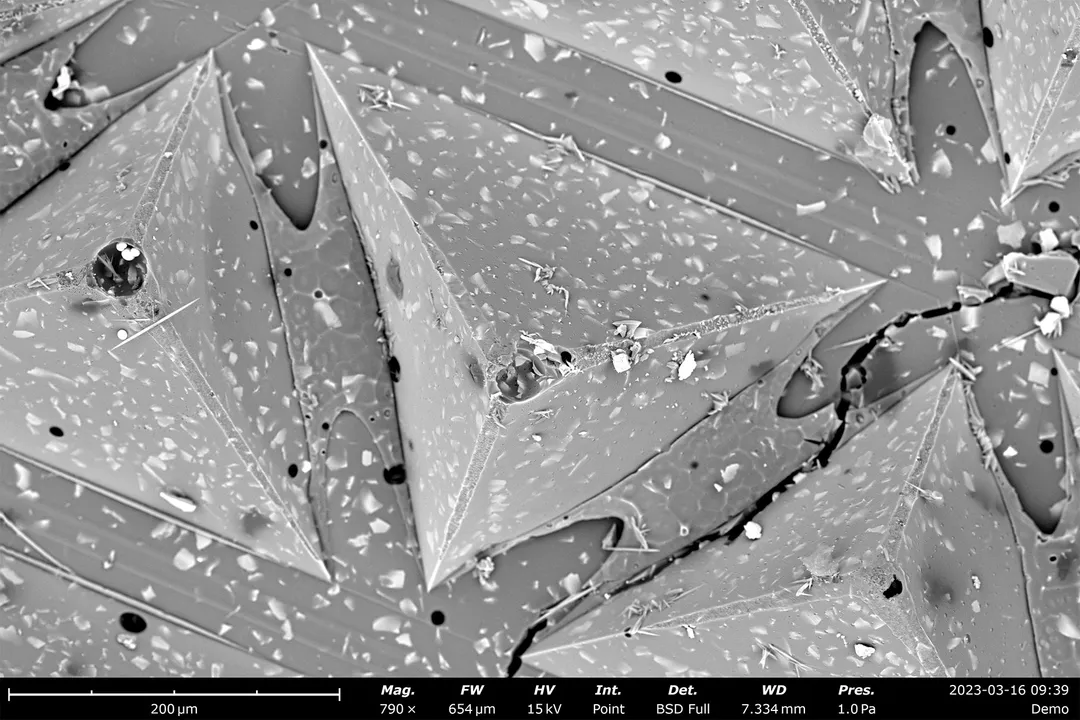
# Surface Versus Internal
What we see through an electron microscope heavily depends on the specific configuration of the instrument being used, primarily splitting into two main approaches: Scanning Electron Microscopy (SEM) and Transmission Electron Microscopy (TEM). [3]
SEM allows us to map the topographical landscape of a sample. [2] The electron beam scans across the surface, and detectors capture secondary electrons emitted from the surface, generating highly detailed, three-dimensional-like images of the exterior. [3][7] This is perfect for examining the texture of a pollen grain, the surface topography of a microchip, or the structure of a fractured material. [7]
TEM, conversely, provides views of the internal architecture. [3] In this setup, the electron beam is transmitted through an ultra-thin slice of the sample. [3] Areas that are denser or absorb more electrons appear darker in the final image, rendering a detailed cross-section of the internal components. [7] For instance, a researcher studying a cell wall might use TEM to examine the layering within the wall, while using SEM to study how that wall interacts with an external bacterium. [2] A practical consideration when choosing is the inherent trade-off: gaining the high internal resolution of TEM often necessitates extensive sample preparation that might obscure subtle surface chemistry visible via SEM.
# Material Structure
Beyond biology, electron microscopy has profoundly impacted materials science and engineering, allowing us to visualize matter at scales relevant to its physical properties. [7] Researchers can observe the arrangement of crystal lattices, analyze defects in metals, and examine the interfaces between different phases within an alloy. [7] Seeing the structure at this level—where imperfections or atomic spacing directly influence strength, conductivity, or flexibility—is indispensable for quality control and discovery. [7] For an engineer analyzing a microscopic fracture in a metal component, the contrast difference between the dense metal structure (seen via TEM) and the surface topography of the break (seen via SEM) provides a complete diagnostic picture far exceeding what lower magnification could offer, guiding material selection or manufacturing process adjustments.

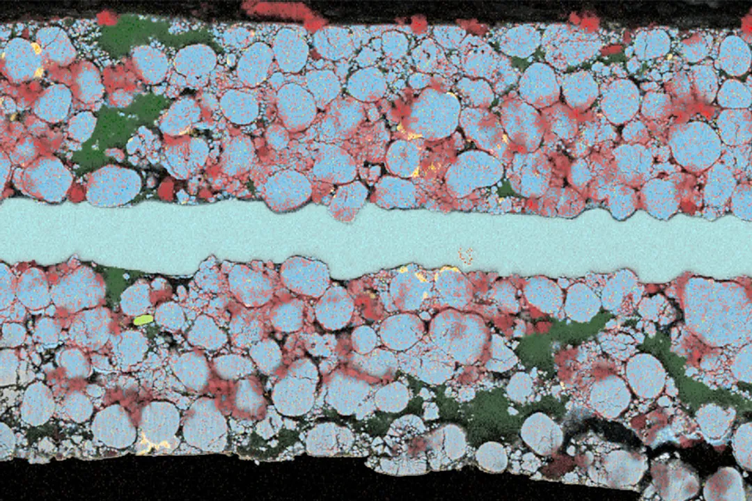
# Property Mapping
The capacity of electron microscopes is extending past simple visualization of form and structure to the analysis of function and composition simultaneously. Certain specialized detectors and techniques integrated into the microscope allow scientists to map out material properties directly correlated with the visual image. [10] For example, techniques like Cathodoluminescence Spectroscopy (CL), when coupled with an electron beam instrument, enable the analysis of the light emitted from a sample as it interacts with the electrons. [10] This emission is directly related to the electronic band structure and chemical composition of the material, particularly useful in semiconductors. [10] Therefore, an electron microscope doesn't just show where something is; advanced configurations reveal what it is made of, or what electrical potential it holds, all within the same instrument chamber. [10] This capability to overlay structural information with compositional data is vital for nanotechnology research, where a few atoms can define a material’s behavior. [7]
In essence, electron microscopes serve as our primary windows into the sub-micron and nanometer worlds. [7] They allow us to see the fundamental components of life, the precise crystalline arrangement of man-made materials, and the subtle interplay between structure and property across scales previously considered unknowable, pushing the boundaries of scientific visualization. [1][6]
Related Questions
#Citations
Electron microscopes - The Learning Zone
What is Electron Microscopy? - UMass Chan Medical School
Electron microscope - Wikipedia
ELI5: How does an electron microscope work? : r/explainlikeimfive
How Does An Electron Microscope Work?
What do electron microscopes allow us to see? - Quora
[PDF] Exploring Uncharted Realms with Electron Microscopy
From Photons to Electrons: Optical and Electron Microscopy
Electron microscopes - Cell structure - Edexcel - BBC
What is an Electron Microscope? - HORIBA