How does ultrasound imaging function?
The technology underpinning ultrasound imaging seems almost like magic: using sound waves to paint a picture of soft tissues deep inside the body without any radiation. It’s a fascinating application of physics, transforming inaudible vibrations into real-time anatomical views, making it indispensable in modern medicine from monitoring a pregnancy to guiding emergency procedures. [1][2][4] The fundamental principle hinges on sending sound pulses into the body and listening carefully for their return, much like a bat uses echolocation, but with much higher precision and specific frequency ranges. [7][8]
# Sound Frequencies
Diagnostic ultrasound operates in a frequency range far above what the human ear can detect. Typical diagnostic frequencies span from about 2 to 18 megahertz (MHz), though some specialized systems can reach up to 50 MHz. [5][8] To put that into perspective, human hearing generally tops out around 20 kilohertz (kHz), or 0.020 MHz. [5] This extreme high-frequency sound is what gives the technique its name—ultrasound. [7] Operating at these high frequencies allows for greater resolution because the shorter wavelengths can distinguish smaller structures. [5] However, there is a trade-off: higher frequencies are absorbed by tissue more quickly, which limits how deep the sound waves can penetrate. [5] Lower frequencies penetrate deeper but provide a less detailed image. [5]
# Transducer Action

The device responsible for both sending and receiving these sound waves is the transducer, often called the probe. [2][6] This small, handheld device is the interface between the machine and the patient. [1] Inside the transducer lies the core component: the piezoelectric crystal. [5][8] These special materials have a unique property; when an electrical voltage is applied across them, they vibrate, or oscillate, producing the high-frequency sound pulses required for imaging. [5]
When the technician presses the probe against the skin, this crystal vibrates rapidly, emitting short bursts of ultrasound energy into the body. [6][7] The process is incredibly fast, involving thousands of pulses per second. [8] As soon as the pulse is sent, the electrical current is turned off, and the crystal instantly switches roles, becoming a sensitive receiver, waiting to detect the returning echoes. [5][7] This ability to instantly switch between generating sound and receiving echoes is fundamental to creating the image. [5]
An interesting design consideration in the physical setup is the need for a coupling medium. Without a conducting substance between the probe and the skin—usually a clear, water-based gel—the sound waves would reflect almost entirely off the air trapped between the probe and the skin surface, as the difference in acoustic impedance between air and tissue is too great. [2][5] This gel ensures the sound energy efficiently enters the body, which is why you always see a generous application of gel before any scan begins. [6]
# Echo Reception
Once the sound pulse enters the body, it travels through tissues until it encounters a boundary between two different types of material—for instance, between fluid and soft tissue, or soft tissue and bone. [1][7] At these interfaces, some of the sound energy is reflected back toward the transducer as an echo, while the rest continues deeper into the body until it hits another boundary or dissipates. [7][8]
The transducer captures these returning echoes. [5] The machine then measures two critical factors for each echo received:
- Intensity (Strength): How strong the returning echo is tells the computer about the tissue interface. Strong echoes come from boundaries where the acoustic properties change dramatically, such as the boundary between liver tissue and fluid-filled cysts or between tissue and bone. [1][7] Weaker echoes come from softer transitions.
- Time of Flight: The time it takes for the echo to return dictates how far away the reflecting structure is from the probe. [5][8] Sound travels at a known, relatively constant speed through soft tissue (approximately 1540 meters per second). [5][8] By multiplying this speed by half the round-trip time, the system precisely calculates the depth of the structure. [5]
The sophistication of the ultrasound system lies in its computer processing unit, which takes these thousands of data points per second—time, intensity, and location—and constructs a two-dimensional visual representation in real-time. [1][8]
# Display Modes
The raw data from the returning echoes must be translated into a visual format the clinician can interpret. Diagnostic ultrasound employs several standard display modes to visualize different aspects of the body's structures and motion. [6][8]
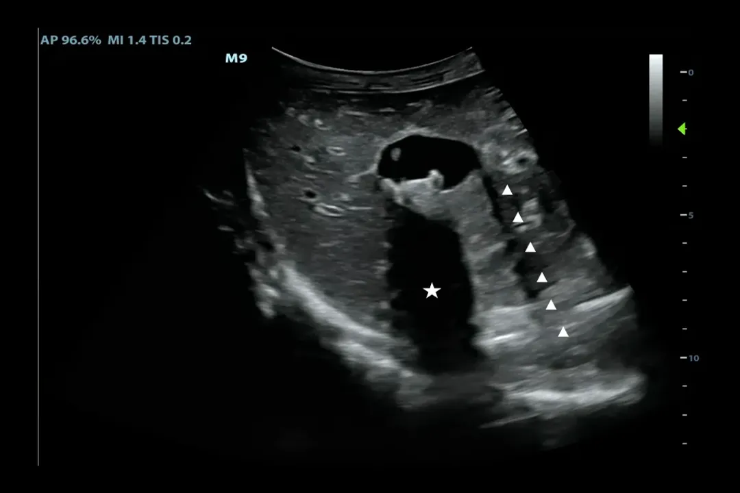
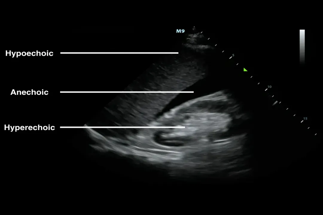
# B-Mode Imaging
The most common format is B-mode, which stands for brightness mode. [6][8] In this mode, the brightness of each dot on the screen corresponds to the intensity of the received echo. [6] A very bright dot indicates a strong reflection (like a solid structure or bone), while a dark or black area indicates that sound passed through unimpeded (like a simple fluid-filled structure, such as a bladder or a simple cyst). [7] B-mode creates the familiar, cross-sectional gray-scale images seen in many diagnostic reports. [6]
# M-Mode Imaging
M-mode, or motion mode, is excellent for studying structures that move over time, such as the heart valves or the fetal heart rate. [6][8] In M-mode, the machine locks onto a single line or beam and records the movement along that line over time. [6] The resulting image displays depth on the vertical axis and time on the horizontal axis, allowing practitioners to precisely measure the rate and character of repetitive motion. [8]
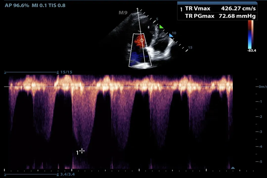
# Doppler Imaging
While B-mode shows anatomy, Doppler ultrasound reveals movement within the anatomy, most importantly blood flow. [4][6] This relies on the Doppler effect—the change in frequency of a wave in relation to an observer who is moving relative to the wave source. [8]
When the sound beam reflects off moving red blood cells, the frequency of the returning echo is slightly higher or lower than the frequency that was sent out, depending on the direction of flow relative to the probe. [5][6]
Doppler is typically displayed in a few ways:
- Color Doppler: Overlays color onto the B-mode image. Red might represent flow moving toward the transducer, while blue represents flow moving away, giving an immediate visual map of circulation paths. [6]
- Pulsed Wave or Continuous Wave Doppler: Provides a spectral display (a graph) of the velocity and direction of flow over time, which is crucial for assessing the severity of blockages or leaks in vessels or heart valves. [5][6]

# Assembling the Image
Modern ultrasound arrays contain hundreds, sometimes thousands, of tiny piezoelectric elements packed tightly together within the probe head. [8] To create a full image, these elements are fired sequentially in precise, timed patterns.
The machine doesn't just fire one beam straight down; it fires beams at slightly different angles, steering the sound electronically to build a sector shape or a wide field of view. [5][8] The computer then correlates the time and angle of return for every echo across this entire field to construct the final, coherent image on the screen. [1][5] This electronic steering is what allows modern scanners to create wide views without needing bulky mechanical parts to pivot the sound beam inside the probe itself. [5]
If you consider the time investment, a standard ultrasound examination, while highly flexible, is often significantly quicker to set up and complete than advanced cross-sectional imaging like an MRI, which can take an hour or more. [4] This speed, coupled with the non-invasive nature, is why ultrasound is favored in emergency settings for quickly assessing trauma or critical conditions. [4]
# Safety and Applications
A major advantage of ultrasound imaging is its safety profile. Unlike X-rays or Computed Tomography (CT) scans, diagnostic ultrasound does not use ionizing radiation. [1][4][7] This is why it is the preferred imaging modality for monitoring the developing fetus throughout pregnancy. [4][7] Concerns about thermal effects (slight heating of tissues from the sound energy) or mechanical effects (the pressure waves) are generally considered minimal with low-power diagnostic settings, adhering to strict safety standards. [1][4]
The technology is remarkably versatile. It can visualize virtually any soft tissue structure, including:
- Obstetrics and gynecology (monitoring pregnancy, examining ovaries and uterus). [2][4]
- Cardiology (echocardiography for the heart). [2][6]
- Abdominal organs (liver, kidneys, gallbladder). [2][4]
- Vascular assessment (checking for clots or narrowing in arteries and veins). [2][6]
- Musculoskeletal imaging (tendons, ligaments, muscles). [4]
While it is unparalleled for soft tissue, its primary limitation remains its inability to penetrate bone or gas effectively, meaning views of the brain (in adults), lungs, or structures deep behind the rib cage can be significantly obscured by these interfaces. [1][7]
When a new practitioner first learns to use a probe, they often find the initial image quality very noisy or indistinct. A key piece of practical advice involves focusing on controlling the gain setting—this is the machine's overall amplification of the returning echoes. Setting the gain too low results in a too-dark image where subtle structures are missed; setting it too high washes out contrast, making everything look gray and blurring the edges between structures. Finding that sweet spot where the background is mostly black but the important anatomical borders are crisp white takes significant hands-on experience. [3][5] This direct relationship between the operator's skill and the resulting image quality highlights the expertise required in sonography that goes beyond simply positioning the probe. [9]
The ability to provide instant, high-resolution information without wires or contrast agents that require separate administration steps gives ultrasound a unique place in point-of-care medicine. Whether guided by an experienced sonographer or used at the bedside by an emergency physician, the simple act of applying the gel and touching the skin opens up a window into the living body. [4][6]
#Videos
How Does Ultrasound Work? - YouTube
Related Questions
#Citations
Ultrasound
Ultrasound: What It Is, Purpose, Procedure & Results
How Does Ultrasound Work? - YouTube
Ultrasound - Mayo Clinic
Ultrasound Physics and Technical Facts for the Beginner - ACEP
Ultrasound (Sonography) Procedures - Radiologyinfo.org
Ultrasound scans: How do they work? - Medical News Today
Medical ultrasound - Wikipedia
Ultrasound principles and instrumentation - ScienceDirect.com