How do neurotransmitters cross synapses?
The communication highway within the nervous system requires an elegant solution for transmitting signals from one specialized cell to the next, especially when those cells do not physically touch. This essential relay occurs at the synapse, a microscopic junction where chemical messengers bridge the gap between neurons. [2][8] Understanding this transfer is key to grasping everything from reflexes to complex thought processes. [1]
# Anatomy Basics
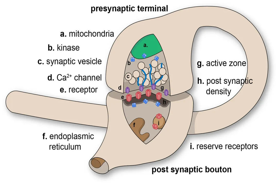
A synapse consists primarily of three parts: the presynaptic terminal (the end of the transmitting neuron), the synaptic cleft (the minute space separating the cells), and the postsynaptic membrane (the receiving surface of the next neuron or target cell). [5][7] While electrical synapses allow ions to flow directly between cells via gap junctions, the process of chemical neurotransmission is far more common and complex. [5] In a chemical synapse, the membranes of the two cells are physically separated by the cleft, which is generally less than 20 nanometers wide. [5][7]
# Action Potential Arrival
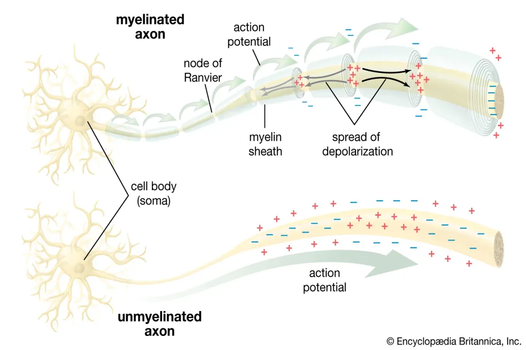
The entire sequence is initiated by an electrical event arriving at the presynaptic terminal. This electrical signal is known as an action potential, which is a rapid, temporary reversal of the electrical charge across the neuron's membrane. [1][6] When this spike of electrical activity reaches the axon terminal, it acts as the essential trigger, signaling that it is time to send a message across the gap. [1]
# Calcium Trigger

The arrival of the action potential does not directly push the chemical messenger across. Instead, the change in voltage causes specialized gates, called voltage-gated calcium channels, embedded in the presynaptic membrane, to fly open. [1][9] Because the concentration of calcium ions () is much higher outside the neuron than inside, these opened channels allow to rush rapidly into the terminal, down its electrochemical gradient. [6][9] This influx of calcium is the non-negotiable prerequisite for neurotransmitter release; without it, the chemical messengers remain locked inside their storage units. [9] A useful way to visualize this is to think of the calcium ions acting as the 'key' that unlocks the 'cargo doors' (the vesicles) of the neuron, whereas the electrical signal itself just got the key to the correct location. [1][9]
# Vesicle Release
Inside the presynaptic terminal, neurotransmitters are packaged into tiny, membrane-bound sacs called vesicles. [7] Once the intracellular calcium concentration rises sufficiently due to the influx, it binds to sensor proteins near the vesicles. [6][9] This binding triggers a cascade of protein interactions that pull the vesicles toward the inner surface of the presynaptic membrane. [9] The vesicle membrane then fuses with the presynaptic membrane—a process called exocytosis—effectively dumping the neurotransmitter cargo into the synaptic cleft. [1][6][7]
# Cleft Crossing
Once the neurotransmitters are released, they face the challenge of crossing the synaptic cleft to reach the receiving cell. They do not travel by active transport or requiring energy for this step; instead, they rely on simple physical principles. [7] The concentration of the neurotransmitter is now extremely high in the cleft, right next to the presynaptic side, and zero on the postsynaptic side. Consequently, the molecules spread out rapidly via diffusion, moving from the area of high concentration to the area of low concentration. [1][7] Because the cleft is so narrow—often less than 20 nanometers—this diffusion process is incredibly fast, allowing the chemical signal to traverse the gap in mere fractions of a second. [5]
Consider the implications of this narrow gap and the concentration gradient. Because the distance is minimal and the gradient is steep, the time spent in the cleft is minimized, often requiring only a fraction of a millisecond. This reliance on rapid diffusion means that the entire structural integrity of the synapse—the exact spacing and the concentration gradient—must be meticulously maintained for normal signaling speed. If the structural organization tethering the vesicles near the membrane were even slightly compromised, the brief delay could accumulate across vast networks, noticeably slowing down complex reflexes or reaction times throughout the central nervous system. [5][7]
# Postsynaptic Binding
The message is only successfully transmitted when the molecules reach the other side. On the postsynaptic membrane, specialized receptor proteins are situated, acting like highly specific locks for the neurotransmitter "keys". [1][2] The neurotransmitter must bind precisely to its matching receptor site to elicit a response. [6]
This binding action causes a change in the postsynaptic receptor, often resulting in the opening or closing of ion channels in that neuron's membrane. [6] Depending on the neurotransmitter and the receptor type, this action will either excite the receiving neuron (making it more likely to fire its own action potential, known as an Excitatory Postsynaptic Potential or EPSP) or inhibit it (making it less likely to fire, an Inhibitory Postsynaptic Potential or IPSP). [6] This is where the initial electrical signal is chemically translated into a specific, graded electrical change in the next cell, which then determines whether that cell continues the electrical wave. [1] Various neurotransmitters, such as acetylcholine, serotonin, or dopamine, have different receptor affinities, leading to the vast array of functions managed by the brain. [4]
# Signal Termination
For the nervous system to function accurately and rapidly, the chemical message must be terminated as soon as it has delivered its instruction. If neurotransmitters remained bound to receptors, the postsynaptic neuron would either be continuously excited or inhibited, preventing the precise timing required for coordinated thought and movement. [1]
There are several primary mechanisms the neuron employs to clear the synaptic cleft rapidly: [6]
- Reuptake: The presynaptic terminal can possess special transporter proteins that actively pump the neurotransmitter molecules back into the releasing neuron, recycling them for future use. [1][6]
- Enzymatic Degradation: Specific enzymes located within the synaptic cleft or on the postsynaptic membrane can chemically break down the neurotransmitter into inactive components. [6] For instance, the enzyme acetylcholinesterase breaks down acetylcholine. [1]
- Diffusion Away: Some molecules simply diffuse away from the active synaptic region, moving into the extracellular fluid where they are diluted to ineffective concentrations. [6]
This rapid "cleanup" process, which relies on the coordinated actions of transporters and degradative enzymes, is just as crucial to fast communication as the initial release mechanism itself. [1] The specific chemicals used for termination can vary significantly between different synapse types, providing another layer of regulatory control over signaling speed and duration. [6]
#Videos
2-Minute Neuroscience: Synaptic Transmission - YouTube
Related Questions
#Citations
Action potentials and synapses - Queensland Brain Institute
Sending Information: Synapses and Neurotransmission - BrainFacts
2-Minute Neuroscience: Synaptic Transmission - YouTube
Neurotransmitters: What They Are, Functions & Types
Synapse - Wikipedia
Synaptic Transmission - Neurotransmission - TeachMePhysiology
Chemical synapses: Structure, function and diagram - Kenhub
Neurotransmission: The Synapse - Dana Foundation
Neurotransmitter release | Description, Synapse, & Process